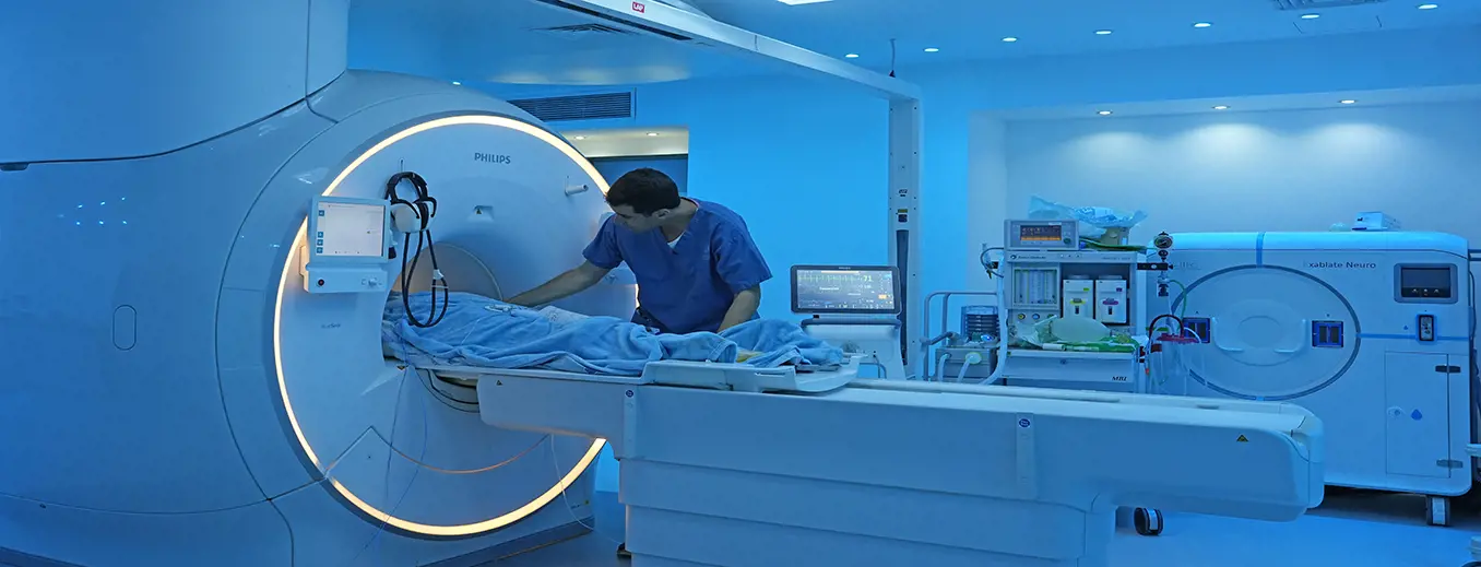
A high-quality imaging examination is critical to the patient's health: A radiologist can be compared to an intelligence officer who prepares charts and maps for a special operation. The more accurate the charts and the more precise the interpretation of the situation, the greater the chances of success for the commander—or, in medicine, the attending physician or surgeon—in the battle to protect the patient's health.
The Imaging Division at Tel Aviv Medical Center is considered one of the largest and best in Israel. The division serves the various departments of the medical center and performs all types of imaging examinations for hospitalized patients and individuals from across the country.
Medical imaging is now the backbone of modern medicine. Physicians from all medical specialties rely on imaging to diagnose patients, assess the effectiveness and suitability of the treatment provided, and determine whether patients are suitable for surgery.
The Imaging Division sets a leading standard in medical imaging services, combining uncompromising professionalism, advanced equipment and technology, and personalized patient care. The goal is to provide patients with the highest-quality imaging to enable optimal diagnosis, treatment, and follow-up for each case.
Team

Useful Information
- Diagnostic Imaging – The Imaging Division performs all types of diagnostic examinations. We are equipped with a large number of imaging devices, including X-ray machines, ultrasound machines, CT scanners, MRI scanners, mammography equipment, X-ray fluoroscopy systems, and more.
- Interventional Imaging – We perform various invasive procedures and treatments under imaging guidance. The division performs all types of biopsies under CT or ultrasound guidance, as well as drainage of abscesses, biliary obstructions, kidney obstructions, and the gallbladder. In the Interventional Radiology Unit and the Interventional Neuroradiology Unit, we also perform catheterization procedures involving various arteries in the chest, limbs, brain, neck, and abdominal organs. Various treatments are also performed for oncology patients.
To schedule an appointment, please send a referral by email to hadmaya@tlvmc.gov.il.
Then call *8801 / 03-6974000, extension 2.
To schedule an appointment online through the appointment scheduling system >>>
Timeframes for receiving imaging examination reports:
-
MRI – Up to 10 business days
-
Ultrasound (US) – Up to 2 business days
-
CT – Up to 7 business days
-
Mammography – Up to 5 business days
Viewing Examination Results in the Personal Area
Imaging examination reports can be viewed in the MybeWell personal area on the Ichilov website.
Alongside the examination report, you can also view the imaging files themselves and share them for viewing (on a desktop computer).
To view results on the website, it is recommended to ensure that your mobile phone number is updated in the system and to have your ID number and an available mobile phone at hand.
Examinations performed in the Imaging Division, such as CT, MRI, ultrasound, and X-ray, can be viewed, downloaded to a personal computer, and shared with a third party.
- If an examination report is available:
Click the "View Imaging" button located next to the examination report document in the examination row. - If an examination report is not available (the examination has not yet been interpreted or does not require a report, such as X-rays):
You can access the entire imaging folder by clicking the "Test Results" folder and then the "Imaging" tab, where you will find a button to view all imaging examinations. - Sharing the imaging folder with a third party:
Click the "Test Results" folder and then the "Imaging" tab, where you will find a button to share the imaging folder.
The examination report is not sent via Israel Post.
Upon completion of the examination, the images will be uploaded to the beWell website, followed by the examination results (the radiology report), in an accessible digital format.
A disc will be provided after the examination upon request only.
Checking the Status of Examination Results
For telephone inquiries regarding the status of examination results from the past month at the MRI, CT, and Nuclear Medicine institutes, call 03-6972041.
Postal Delivery and Disc Copies
Examination reports are sent via Israel Post to the address provided at the time of the examination. The report will be mailed to the residential address listed in the records only if you requested this during registration for the examination.
Copies of imaging examination discs can be obtained on any working day during business hours.
Examination results and copies of images can be obtained at the X-ray Institute in Wing E, Station No. 4.
Requesting a Disc Copy
In person: Sunday–Thursday, 8:00–14:00. Waiting time is up to two hours. Please bring an ID card. If a representative is collecting the disc on behalf of the patient, a power of attorney is also required.
Without visiting in person: To obtain additional disc copies of imaging examinations, please send a copy of your ID card, a power of attorney, and a contact phone number by fax to 074-7219818 or by email to XRAY-c@tlvmc.gov.il. We will contact you within three business days to arrange payment by credit card over the phone.
The disc will be sent by mail or may be collected in person from the X-ray Institute, Sunday–Thursday.
Eligibility for Receiving a Disc
Obtaining a copy of an examination on disc involves a fee. The cost of a disc copy varies according to the Ministry of Health price list.
Outpatients: Patients who undergo an ultrasound, MRI, CT, mammography, or angiography examination on an outpatient basis are entitled to receive their first copy of the images free of charge, either at the time of the examination or by mail.
Emergency Department / Hospitalized Patients: Patients who undergo an imaging examination in the Emergency Department or during hospitalization are entitled to receive a disc copy at the applicable rate on that date. The cost varies according to the Ministry of Health price list.
Patients requesting copies of several imaging examinations stored in the system, requiring more than one disc, will be charged the price of two discs.
Exceptions:
-
Soldiers performing mandatory military service – Are entitled to receive copies of imaging examinations free of charge upon presentation of a valid military ID card.
-
Attorneys – An attorney requesting a disc copy on behalf of a client will be referred to the Medical Records Department, through which the required discs will be provided.
Before an appointment at Ichilov for an imaging examination or at one of the various specialist clinics, you will be required to bring imaging examinations. If these examinations were not performed at Ichilov, it is recommended to upload them to the Ichilov systems before your appointment.
Using the imaging disc upload system, you can upload your examination in two ways:
1. Remotely – From home, via the following link: Access the Disc Upload System
2. At the hospital – Using self-service kiosks located in the hospital clinics.
Uploading imaging examinations to the system in advance provides a complete picture, enabling optimal medical care.
Inquiries / Questions After Receiving the Report
If you have a question or issue relating solely to the interpretation of your examination:
You can access the following link >>>
Or scan the QR code:

Operational and Professional Imaging Units
The Ultrasound Unit performs ultrasound examinations, including invasive procedures and Doppler examinations as needed, for adults, children, and newborns. The unit is equipped with some of the most advanced technology of its kind in the world and performs a wide range of examinations.
Ultrasound is one of the oldest imaging methods available. It is a relatively quick examination that does not use ionizing radiation and is therefore not harmful to health. It was first introduced as a diagnostic examination in the middle of the last century. The examination is based on the use of ultrasound waves. The ultrasound device generates a wave of a specific frequency and wavelength, which is transmitted to the body's organs through a transducer and a liquid medium (gel). The transmitted wave passes through the organ's tissue and returns to the transducer after undergoing changes within the tissue. The changes in the returning wave are converted into a digital image displayed on the device's screen. This makes it possible to assess the texture and structure of the tissue and identify anatomical changes.
Because ultrasound is readily available, portable, inexpensive, and does not involve radiation, it serves as a basic first-line diagnostic tool for evaluating common conditions such as leg swelling, abdominal pain, urinary tract problems, and more. It helps determine whether a pathological condition exists and where it is located, and often enables effective diagnosis of the problem.
Diagnostic accuracy is the first and most important step toward appropriate treatment. The Ultrasound Unit at Ichilov Hospital offers a unique combination of expertise, technology, extensive experience, and personalized care.
The unit has a multidisciplinary team consisting of radiologists, certified radiographers, service and reception representatives, and a dedicated nursing team. The team accompanies patients throughout the entire process, from appointment scheduling and patient admission to the examination itself and receipt of the radiology report.
The Computed Tomography (CT) Unit is a national leader in its field, performing examinations for patients of all ages (including examinations under general anesthesia when required) and for all parts of the body, including biopsies and drainage procedures. The unit provides services to the entire hospital, including Emergency Department patients, hospitalized patients, and outpatients.
The unit is equipped with the most advanced CT scanners and continuously upgrades its equipment in accordance with global developments in the field, aiming to provide patients with the highest-quality CT examinations while using the lowest possible radiation doses.
The examinations are interpreted by some of Israel's leading radiologists and performed by experienced imaging technologists who specialize in the field.
The unit's specialist radiologists have extensive experience in interpreting examinations across a wide range of diseases. Most have completed advanced training and fellowships at leading hospitals in the United States, Canada, and France, with specialized expertise in all areas of imaging, including brain and nervous system imaging, cardiac and pulmonary imaging, abdominal and pelvic imaging, musculoskeletal imaging, vascular imaging, breast imaging, and pediatric imaging.
The Wahl Magnetic Resonance Imaging (MRI) Institute at Ichilov Hospital, Tel Aviv Medical Center, is one of the largest and most advanced MRI institutes in Israel and ranks alongside leading MRI centers worldwide.
The institute is equipped with some of the newest and most advanced MRI scanners in the world and has a large, experienced team of specialists and service representatives who make every effort to provide patients with high-quality, efficient, and optimal medical care.
The institute's team includes experienced radiologists specializing in a wide range of fields and subspecialties, many of whom have completed advanced training abroad. Alongside the physicians is a skilled multidisciplinary team of radiographers, nurses, physicians, paramedics, and service representatives, who accompany patients from appointment scheduling, through admission and the examination itself, to receipt of the radiology report.
The MRI (Magnetic Resonance Imaging) scanner is a device based on magnetic resonance technology that provides high-quality, detailed images of the body's internal structures without the risks associated with ionizing radiation, enabling early diagnosis and appropriate, precise medical treatment.
The institute has four new, advanced MRI scanners manufactured by Siemens. Assignment to a specific scanner is determined according to medical suitability and clinical considerations.
Types of MRI Examinations
-
MRI of the nervous system: Brain and spine.
-
MRI of the musculoskeletal system: Joints and bones, including MR arthrography (imaging of joints following the injection of contrast material into the joint).
-
MRI of the chest and heart.
-
MRI of the abdomen: Including imaging of the bile ducts (MRCP), bowel enterography (MRE), and urinary tract – MR urography (MRU).
-
MRI of the pelvis: Reproductive system and prostate.
-
Breast MRI: Including biopsies.
-
MRI of blood vessels: MR angiography.
-
Pediatric MRI: Examinations for children under anesthesia.
-
Pediatric MRI without anesthesia.
-
MRI examinations for the detection of fetal abnormalities.
-
In addition, advanced functional imaging examinations are performed as part of the Center for Brain Function Research (fMRI).
Receiving MRI Examination Results
MRI examinations are interpreted within 10 business days, starting from the day following the examination, and the results are displayed in the beWell medical record on the Ichilov website.
Results can be viewed using an ID number, date of birth, and a one-time verification code.
Important! When scheduling your appointment and upon arrival for the examination, please ensure that your personal details in the hospital's records are up to date.
The beWell medical record provides access to the imaging files themselves, including the option to share them with a third party.
1. Access through Tests > Imaging: Clicking the "All Imaging Examinations" button opens the imaging system, where all imaging examinations performed at Ichilov are displayed.
2. Access through Tests > Imaging: Clicking the "View Imaging" button next to the radiology report takes you directly to the relevant imaging examination.
The radiology report is not sent via Israel Post.
Upon completion of the examination, the images will be uploaded to the beWell website, followed by the examination results (the radiology report), in an accessible digital format.
A disc will be provided after the examination upon request only.
The unit performs examinations for infants, children, and adolescents, conducted by a team of radiographers and radiologists with extensive skills, experience, and expertise in pediatric imaging. The examinations are performed using advanced ultrasound equipment and innovative techniques, including examinations with sonographic contrast agents.
The unit serves as a training and educational center for radiologists and radiographers from across the country, as well as residents and students.
Ultrasound is an examination used to visualize various organs of the body using sound waves. The method does not involve exposure to ionizing radiation, which is a clear advantage when examining fetuses, infants, and children, who are particularly sensitive to the harmful effects of radiation. In addition, visualization of internal organs in infants and children is better than in adults due to their smaller body size.
The examination is painless and non-invasive. Parents, including pregnant women, may remain alongside their child during the examination.
Examinations are performed on an urgent basis (through the Emergency Department or during hospitalization) or as outpatient examinations, according to the clinical indication.
Types of Examinations
-
Abdominal / Urinary Tract Ultrasound
-
Ultrasound-Guided Voiding Cystourethrography (CeVUS) in Children
-
Ultrasound for Intestinal Malrotation
-
Spinal Ultrasound
-
Brain Ultrasound
-
Neck and Soft Tissue Ultrasound
-
Scrotal Doppler Ultrasound
-
Transabdominal Pelvic Ultrasound in Girls
-
Contrast-Enhanced Ultrasound with Intravenous Contrast Agent
The Interventional Oncology Unit is part of the Interventional Radiology Unit and specializes in minimally invasive treatments for malignant and benign tumors. Its goal is to provide alternatives to traditional treatments such as surgery or radiation therapy, while minimizing damage to healthy tissues and improving patients' quality of life.
Areas of Activity of the Interventional Oncology Unit
-
Tumor Ablation – Destruction of tumors using heat (radiofrequency or microwave) or freezing (cryoablation).
-
Chemoembolization (TACE) – Injection of chemotherapy directly into the blood vessels supplying the tumor, followed by their blockage to prevent blood supply.
-
Radioembolization (TARE) – Treatment of liver tumors by injecting radioactive particles directly into the tumor's blood vessels.
-
Image-Guided Biopsies – Collection of tissue samples from tumors using a needle, without the need for surgery.
-
Treatment of Chronic Pain Caused by Neuropathy – Targeted injection into the affected nerve area under CT guidance.
Advantages of Interventional Oncology Techniques
-
Less invasive than surgery – Faster recovery and fewer complications.
-
Targeted treatments – Minimal damage to healthy tissues.
-
Can be combined with other oncological treatments – To enhance effectiveness.
The unit works in close collaboration with oncologists, surgeons, and other specialists, accompanying patients throughout all stages of treatment and follow-up.
The treatments are available to patients from across Israel and around the world who come to us to receive advanced care. Each physician in the unit operates a dedicated clinic for appointment scheduling and personal consultations.
The field of medical imaging includes a variety of essential techniques – X-ray, ultrasound, computed tomography (CT), magnetic resonance imaging (MRI), and more – and serves as a key diagnostic tool for physicians in every medical department.
In the past, physicians had to rely solely on their senses and sometimes perform unnecessary surgeries. Today, thanks to imaging technologies, it is possible to look inside the human body and obtain immediate, reliable, and accurate information for diagnosis and treatment – all with the help of medical imaging technologists.
Medical imaging technologists are the professionals who bridge the gap between medical knowledge and advanced imaging equipment. Thanks to their professional training and clinical experience, they perform high-quality examinations that provide physicians with the most accurate diagnostic information and help save lives.
The School of Medical Imaging at Ichilov Medical Center offers advanced professional training that combines theoretical studies with hands-on clinical experience from the early stages of the program. Students benefit from access to innovative imaging equipment, close guidance from experienced professional teams, and a multidisciplinary learning environment at one of Israel's leading university hospitals. With over 60 years of experience in training medical imaging technologists, the school maintains close ties with the healthcare system, helping its graduates quickly enter the workforce in hospitals, imaging institutes, and research centers in Israel and around the world.
The School of Medical Imaging at Tel Aviv Sourasky Medical Center (Ichilov) operates in collaboration with Bar-Ilan University and offers an integrated practical and academic study program:
Bachelor of Science in Medical Imaging (B.Sc.) – Joint Program with Bar-Ilan University
This is a structured bachelor's degree program in medical imaging that provides both an in-depth scientific foundation and comprehensive clinical training.
The school also offers a unique career conversion program for university graduates seeking to enter the field of medical imaging.
When it comes to diagnosing and monitoring cancer patients, accuracy and attention to the smallest details can make all the difference.
The Oncologic Imaging Service at Ichilov Hospital provides in-depth, advanced interpretation of CT and MRI examinations, combining extensive medical knowledge with a unique understanding of oncological treatment.
Our service is led by a leading specialist in the field with over a decade of experience, including specialized training at a prestigious cancer center in New York. It is based on close collaboration with leading oncologists and surgeons, as well as physicians from other disciplines as needed. This includes joint meetings and weekly clinical discussions to provide patients with accurate radiological consultation and the personalized, tailored care they require.
Our service provides advanced interpretation of CT, MRI, and PET-CT examinations, with an emphasis on maximum accuracy and a broad clinical perspective. We specialize in the early detection of tumors, assessment of response to oncological treatments, detection of disease recurrence, and differentiation between malignant and benign changes.
In addition, we are available to provide consultation and guidance to the treating medical team, including in-depth discussions of imaging findings, consideration of treatment options based on imaging results when necessary, and coordination between different medical specialties to support the best possible decision-making for each patient.
The Body Imaging – Abdomen and Pelvis Unit is the largest of its kind in Israel, with approximately 20 radiologists specializing in body imaging, and is a leading unit in the field nationwide. The unit offers advanced diagnostic and interventional therapeutic services, combining extensive medical knowledge, innovative technologies, and a comprehensive clinical approach.
The Body Imaging – Abdomen and Pelvis Unit specializes in imaging the abdominal and pelvic organs, including the liver, pancreas, spleen, kidneys, gastrointestinal tract, urinary system, uterus and ovaries, prostate, and other tissues.
Thanks to its professional team, advanced equipment, and multidisciplinary approach, the unit plays a critical role in accurate diagnosis, developing personalized treatment plans, monitoring complex diseases, assessing patients' suitability for surgery and complex treatments, and improving patients' quality of life.
The unit specializes in both diagnostic and interventional imaging, performing a wide range of complex procedures under the guidance of various imaging modalities.
The unit is equipped with a wide range of imaging devices, all of which are new and among the most advanced in Israel and worldwide.
Tel Aviv Medical Center is at the forefront of chest and cardiac imaging, utilizing advanced technologies and multidisciplinary teamwork to provide the highest quality of care.
The Chest and Cardiac Imaging Unit consists of a dedicated team of four radiologists and two cardiologists, offering a unique combination of expertise. The radiologists, highly skilled in advanced CT and MRI examinations, work closely with cardiologists who combine extensive clinical experience with subspecialty training in cardiac imaging. This multidisciplinary collaboration ensures high-quality care, from accurate diagnosis to treatment planning and long-term follow-up, establishing the unit as a leading, focused service that provides personalized medical care tailored to each patient.
The unit's unique multidisciplinary and collaborative approach, its professional team, and state-of-the-art technology enable imaging-based decision-making and optimal management of a wide range of conditions. These include critical, life-threatening situations in which timely intervention can save lives, as well as preventive medicine aimed at early detection and prevention of the progression of coronary artery atherosclerosis. The upcoming installation of a revolutionary CT scanner will ensure that Tel Aviv Medical Center continues to lead in excellence in chest and cardiac imaging services.
Examinations Performed by the Unit
-
Chest X-rays
-
Chest CT examinations (including pulmonary CT angiography and high-resolution CT of the lungs)
-
Low-dose chest CT examinations (including screening for early detection of lung cancer)
-
CT-guided biopsies of lung lesions
-
Cardiac CT angiography (virtual cardiac catheterization)
-
Cardiac and chest MRI examinations
Breast imaging consists of two main areas: diagnostic imaging and interventional procedures.
Traditionally, interventional procedures performed by radiologists have primarily involved image-guided biopsies of breast lesions. The purpose of these biopsies is to obtain a tissue diagnosis and differentiate between benign and malignant findings.
In recent years, the role of radiologists in breast interventions has evolved. Beyond their traditional role of performing diagnostic biopsies, radiologists are increasingly taking on a therapeutic role. In many cases, surgery can be avoided by removing lesions through percutaneous, image-guided procedures, thereby preventing complications associated with open surgery and improving cosmetic outcomes for patients.
The Breast Minimally Invasive Procedures Unit provides two main services:
-
Vacuum-Assisted Percutaneous Excision of High-Risk Breast Lesions
-
Image-Guided Cryoablation of Malignant Breast Tumors
The Breast Imaging Unit at Tel Aviv Medical Center (Ichilov) performs a comprehensive range of the most advanced breast imaging examinations. The unit has a skilled and experienced team, including reception and service representatives, radiographers, and highly experienced radiologists specializing in breast imaging.
The unit operates in close collaboration with breast imaging specialists and experts from various fields of breast health, including oncology, surgery, plastic and reconstructive surgery, pathology, and other specialties, with the aim of providing comprehensive, personalized care for every patient.
The breast imaging services provided by the unit address the full range of medical needs, from screening examinations for early detection and diagnosis, through routine examinations, to follow-up care and advanced treatments performed in the Breast Minimally Invasive Procedures Unit.
Breast Imaging Examinations and Procedures Performed in the Unit
-
Mammography
-
Breast Biopsies
-
Breast Ultrasound
-
Advanced Breast Imaging Examinations – MRI and More
-
Minimally Invasive Procedures
The unit is part of the Imaging Division at Tel Aviv Medical Center and performs a wide range of imaging and diagnostic examinations for toddlers, children, adolescents, and pregnant women, ranging from fluoroscopy and X-ray examinations to low-dose CT scans and ultrasound examinations, as well as complex MRI examinations under general anesthesia.
The Pediatric Imaging Unit serves as a center of excellence, providing the full range of diagnostic imaging examinations for children, with planning and coordination of the various examinations according to the child's age and clinical condition.
The unit provides comprehensive, integrated care for structures of the musculoskeletal system, including bones, joints, and soft tissues.
The unit specializes in imaging trauma-related conditions, joint diseases, tumors of the musculoskeletal system and soft tissues, and various metabolic diseases.
The unit utilizes all imaging modalities, including conventional X-rays, CT, MRI, and ultrasound.
Thousands of CT, MRI, and ultrasound examinations are performed and interpreted annually in the unit, for both hospitalized patients and outpatients.
The unit also performs interventional procedures, including CT- and ultrasound-guided biopsies of musculoskeletal and soft tissue lesions (more than 400 annually), shoulder and hip arthrography, and injections into various joints.
The unit operates through collaboration within the department and close cooperation with other hospital departments, including the National Orthopedic Oncology Department, the Rheumatology Department, adult and pediatric orthopedic departments, and other hospital departments.
The uniqueness of this unit lies in its comprehensive approach to patients' medical conditions, with an emphasis on neurological disorders and their possible connection to other body systems. The unit works in full collaboration with the Neurosurgery and Neurology Divisions, as well as all hospital departments treating patients with neurological conditions.
Diagnostic Neuroradiology
Imaging of the central nervous system, including the head, neck, and spine. Imaging is performed using CT, MRI, angiography, and myelography, for both hospitalized patients and outpatients.
The uniqueness of this unit lies in its comprehensive approach to patients' medical conditions, with an emphasis on neurological disorders and their possible connection to other body systems. The unit works in full collaboration with the Neurosurgery and Neurology Divisions, as well as all hospital departments treating patients with neurological conditions.
Cerebral Catheterization – Interventional Neuroradiology
The unit specializes in the diagnosis and treatment of vascular disorders of the brain, face, and spine, using groundbreaking technologies.
The field of neuroradiology emerged in Europe in the mid-1960s and has gained momentum over the past decade due to technological advances in medical equipment and imaging devices, such as MRI and CT. These advances have significantly improved the ability to treat vascular disorders and diseases of the brain and central nervous system, including stroke, cerebral aneurysms, and cancerous tumors.
These treatments are performed with maximum precision and minimal complications, reflecting the current trend, whenever the nature of the condition allows, toward preferring minimally invasive catheter-based interventions over more extensive surgical procedures.
In addition to significant advances in imaging technologies, new medical devices have been developed specifically for treating vascular disorders of the nervous system, including coils, stents, various types of medical adhesives, and more.
Thanks to these developments, medical teams with specialized expertise in these fields can safely access the blood vessels of the neck, brain, and spinal cord using miniature instruments to treat various vascular conditions.
Endovascular Interventional Neuroradiology Is Suitable for the Following Conditions and Procedures:
-
Acute ischemic stroke – Reopening blocked arteries during the acute phase of a stroke.
-
Cerebral aneurysms.
-
Arteriovenous and venous malformations.
-
Fistulas.
-
Significant stenosis of cervical and intracranial blood vessels.
-
Tumor embolization – Occlusion of blood vessels supplying tumors as a preliminary treatment before surgery for highly vascular tumors.
-
Cerebral functional testing – Performing tests to identify the location of functional brain areas.
-
Direct alcohol injections – Injection of alcohol (which destroys tissue) into lesions in the spine, face, and other organs.
-
Diagnosis and treatment of spontaneous intracranial hypotension (SIH) – Performing Digital Subtraction Myelography (DSM) to identify cerebrospinal fluid–venous fistulas and direct cerebrospinal fluid leaks, as well as CT myelography. Endovascular treatment is also performed to close cerebrospinal fluid–venous fistulas identified using these diagnostic methods.
The Sagol Brain Institute was established at Tel Aviv Sourasky Medical Center in 1998, in collaboration with Tel Aviv University. The Institute integrates extensive research activities with clinical services.
The Institute's activities are dedicated to studying human brain function by characterizing multidimensional information on the brain's structure and activity, using advanced neuroimaging techniques, including MRI and EEG.
The Institute is directed by Prof. Yuval Nir and was founded by Prof. Talma Hendler, a psychiatrist and neuroscientist, together with Associate Director Prof. Daphne Ben Bashat, an expert in MRI-based neuroimaging.
Clinical Activity at the Institute
Alongside its research activities, the Institute is committed to providing advanced solutions for precise and detailed brain assessment, as well as neuromodulation-based therapeutic approaches for a wide range of brain disorders. These services are provided through the Advanced Functional Mapping Service and the Neuromodulation Clinic.
Psychedelic Research Program
A Joint Initiative of the Institute for Psychedelic Research at Tel Aviv University and the Sagol Brain Institute
The Psychedelic Research Program, led by Prof. Talma Hendler, together with Dr. Nimrod Keynan (Scientific Manager) of Tel Aviv University and Dr. Haggai Sharon (Medical Director) of Ichilov, was established to deepen scientific and clinical understanding of the safe and informed medical use of psychedelic compounds.
The program focuses on patients suffering from treatment-resistant neuropsychiatric disorders, including post-traumatic stress disorder (PTSD), depression, chronic pain, and addictions. It also investigates fundamental mechanisms of the psychedelic experience in healthy individuals and its effects on learning, emotions, and cognition.
Several studies are currently underway as part of this program.
The institute performs examinations to assess various body systems using X-ray imaging, including chest X-rays, abdominal and pelvic X-rays, orthopedic X-rays of the bones, and fluoroscopic examinations to evaluate the urinary and digestive systems (esophageal and gastric fluoroscopy, barium swallow examinations, and imaging examinations prior to obesity treatment surgery (bariatric surgery)). Various examinations are also performed for children.
Appointment Scheduling and Obtaining Disc Copies
- X-ray examinations for adults, excluding fluoroscopic examinations and VCUG examinations, do not require an appointment. Patients may arrive Sunday through Thursday, between 8:00 and 13:00, with a medical referral and a payment authorization form or payment.
- For inquiries and appointment scheduling for fluoroscopic X-ray examinations, please send a referral letter from your physician by fax to 074-7219818 or by email to XRAY-c@tlvmc.gov.il. We will contact you within 3 business days.
- To obtain additional disc copies of imaging examinations, please send a copy of your ID card, a power of attorney, and a contact phone number by fax to 074-7219818 or by email to XRAY-c@tlvmc.gov.il. We will contact you within 3 business days.
The Neuroradiology Unit specializes in imaging the central nervous system, including the head, neck, and spine.
The field of neuroradiology focuses on imaging the nervous system and diagnosing related medical conditions. From back pain and epilepsy to brain tumors, the field covers a wide range of conditions and plays a critical role in determining the diagnosis and treatment of various pathologies, including degenerative, traumatic, neoplastic, vascular, and infectious disorders.
Neuroradiological imaging is primarily three-dimensional and is performed using advanced technologies such as CT (Computed Tomography), which uses ionizing radiation, and MRI (Magnetic Resonance Imaging), which uses electromagnetic energy. Brain and spine MRI examinations account for the majority of MRI scanner utilization.
At the Interventional Radiology Unit (Angio), we perform minimally invasive, image-guided treatments that enable more precise, faster, and safer medical procedures – without the need for complex surgeries. The benefits include shorter recovery times, fewer complications, less pain, and a reduced need for prolonged hospitalization. We are proud to lead the field in the use of advanced technologies and innovative treatment methods.
The unit has three advanced catheterization suites, including a unique integrated CT system (NEXARIS) – the first of its kind in Israel! This system fully integrates interventional CT with fluoroscopy, enabling complex procedures to be performed with maximum precision while reducing procedure time and radiation exposure. We also use several portable ultrasound devices that enable procedures to be performed using innovative techniques.
The unit works in close collaboration with leading departments at the medical center, including:
-
Vascular Surgery Department
-
Oncology Division
-
Liver Surgery and Organ Transplantation
-
Gastroenterology and Nephrology
The medical team actively participates in multidisciplinary meetings to ensure the best possible medical approach for each patient.
The unit not only performs treatments but also monitors patients' conditions following procedures to ensure treatment effectiveness and efficiency. Close follow-up enables optimal care and early identification of any need for additional intervention.