Ichilov
Tel Aviv Sourasky Medical Center

Lis Maternity and Women's Hospital
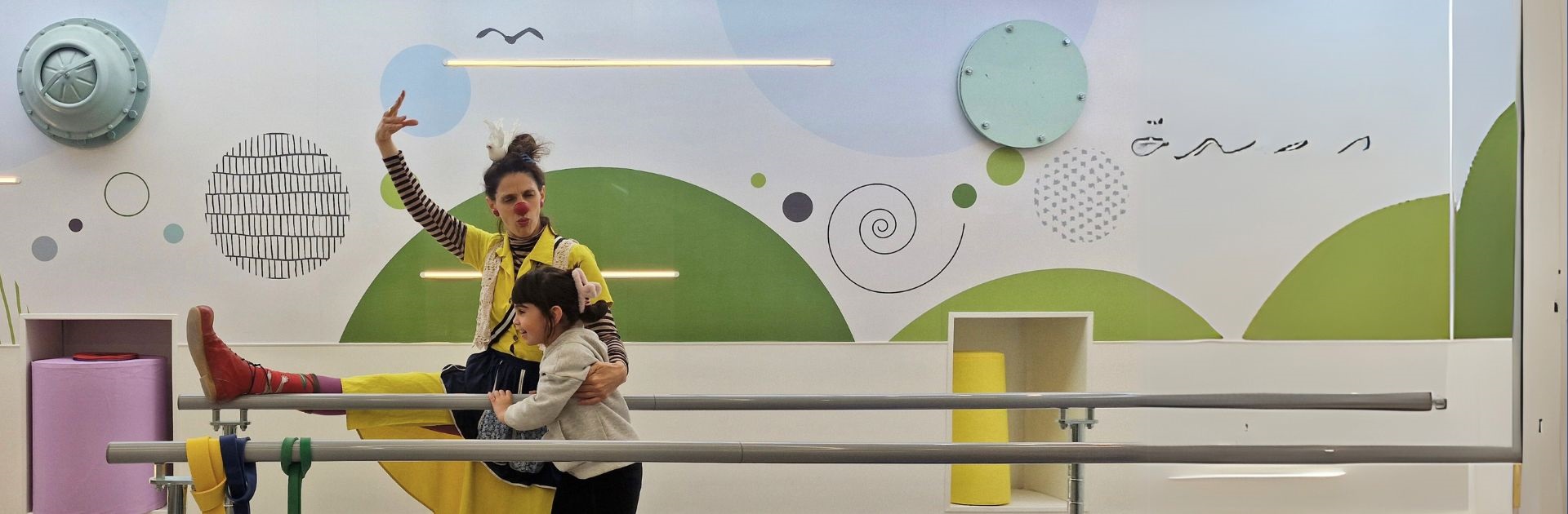
Dana-Dwek Children’s Hospital
About Tel Aviv Sourasky Medical Center
-
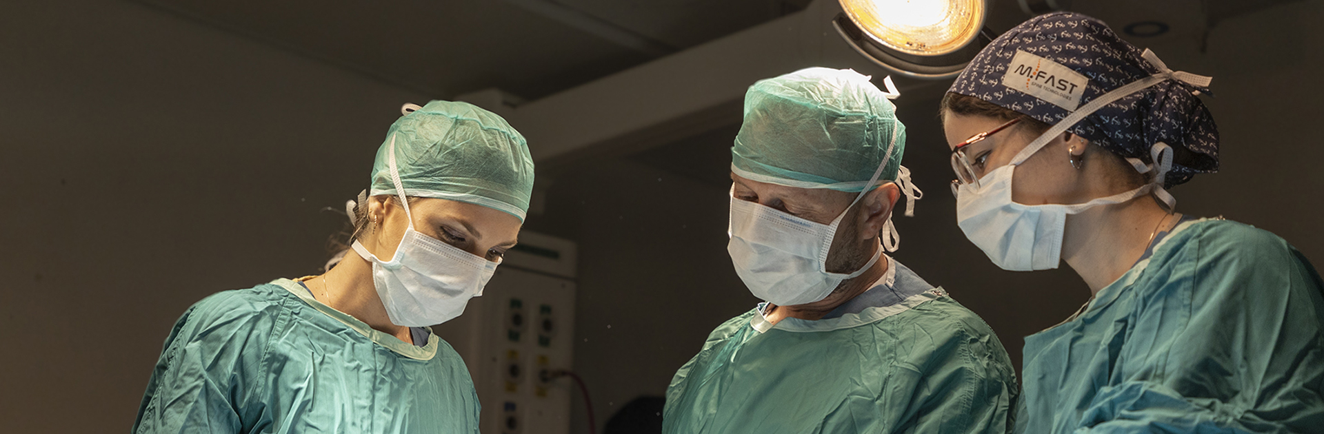
The largest acute care facility in Israel
A 1500-bed world-class governmental academic medical center, Tel Aviv Sourasky Medical Center serves a population of one million people, including residents from the greater Tel Aviv area and visitors to the metropolis. -
Vision for patient-centered care
Tel Aviv Sourasky Medical Center is committed to providing medical excellence and compassionate care, delivered by the most skilled physicians and medical teams, treating each patient equally, and integrating excellence in clinical care -
At the forefront – nationally and internationally
well-deserved reputation as a national referral center across a range of medical and surgical disciplines.
Divisions:
Tel Aviv Sourasky Medical Center patient activity
Patient births since the beginning of the year
Twins were born with us
Girls were born with us
Boys were born with us
Outpatient clinics
Hospitalizations
Surgeries
Emergency Department