Our Vision
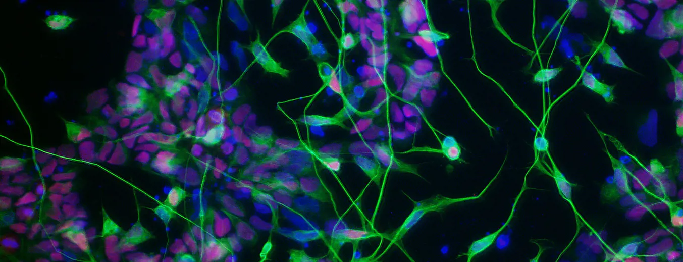
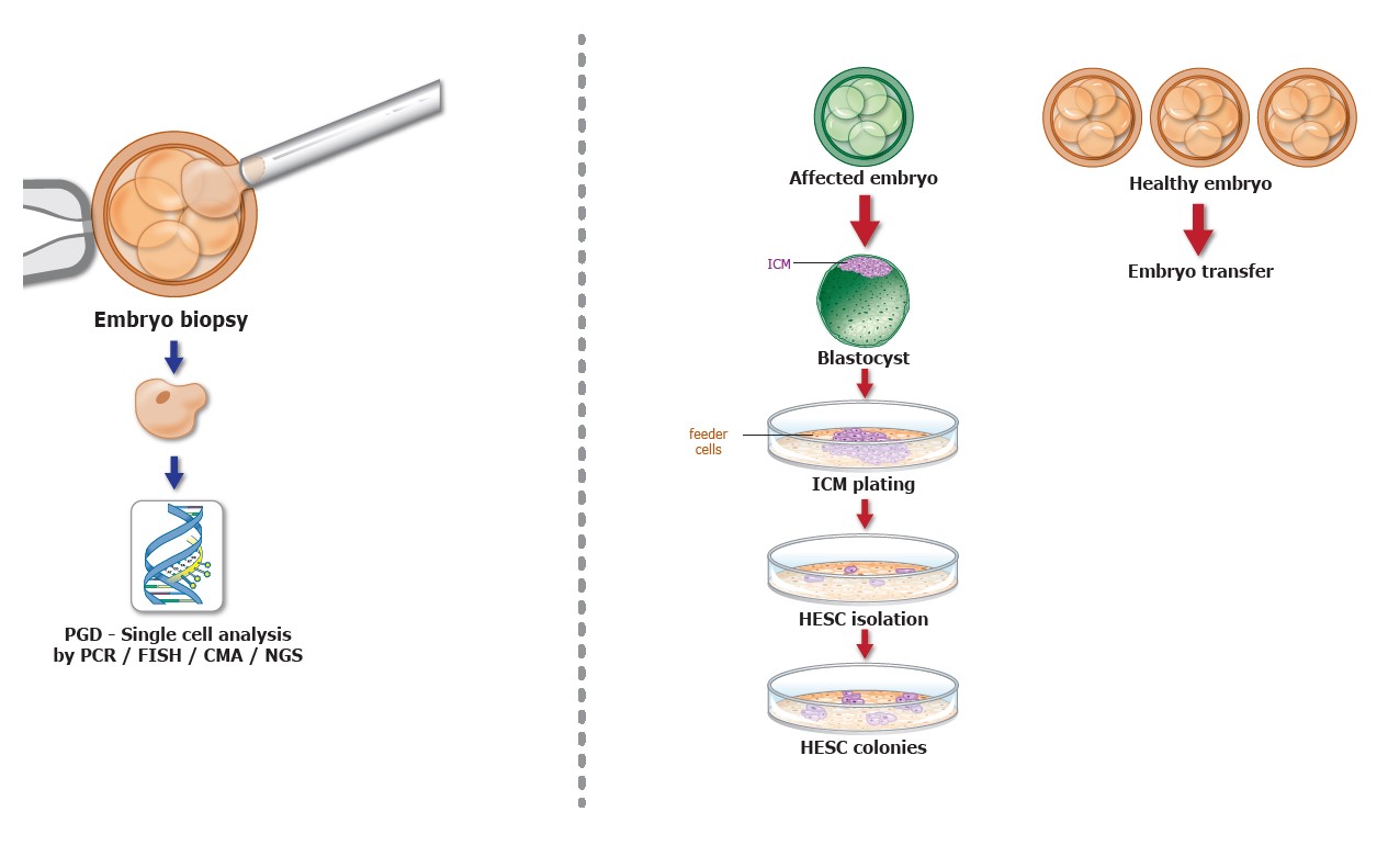
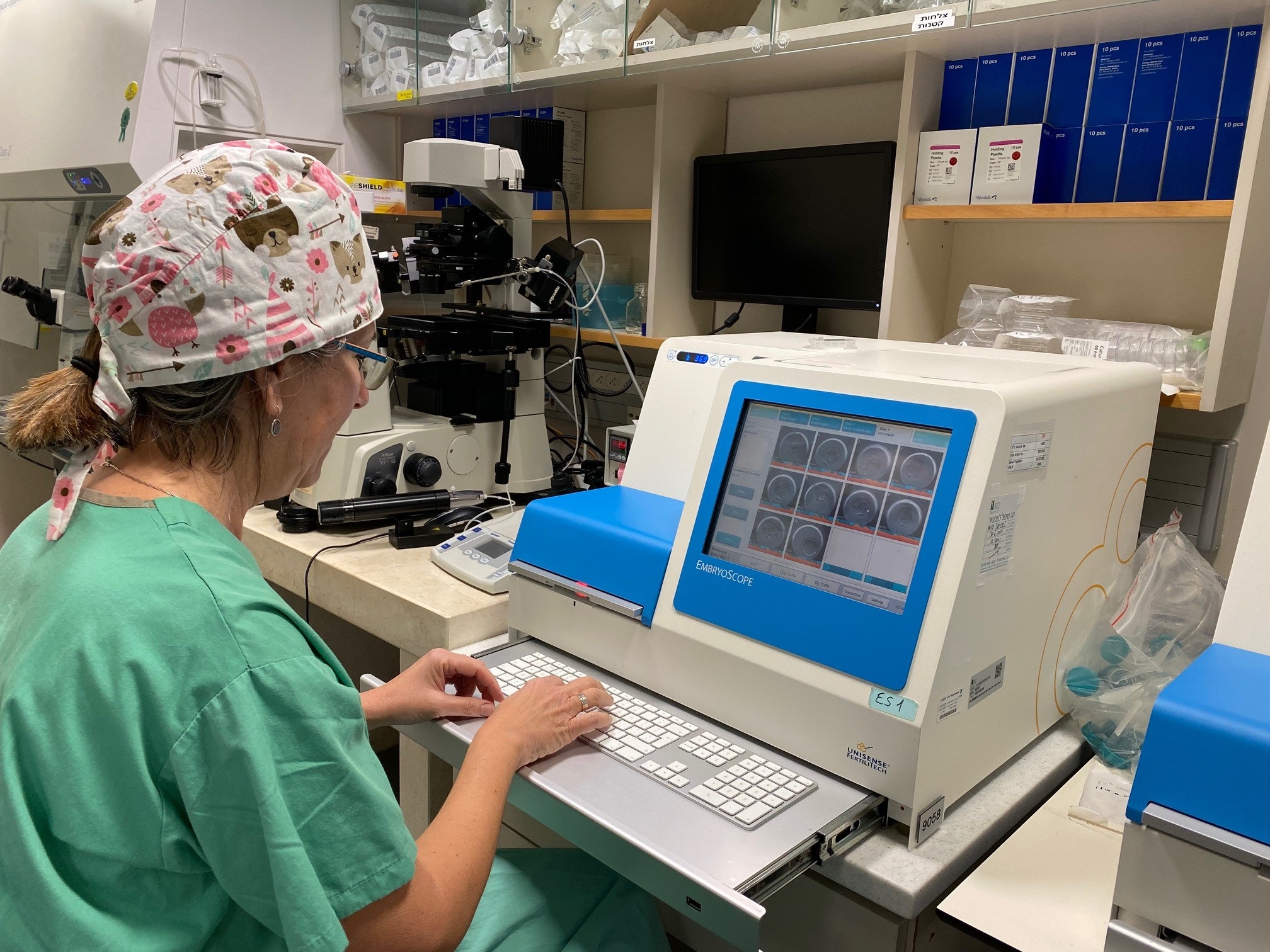
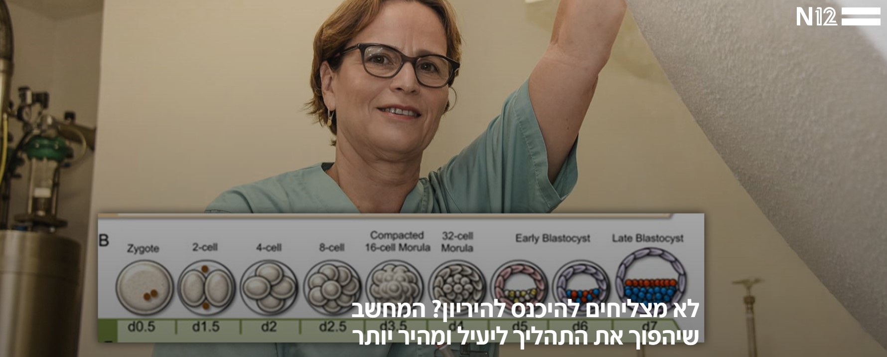
During fertilization, the human sperm and egg unite to form the developing fetus. These early phases of preimplantation development are considered one of the most fundamental questions in cell biology. Our research lab focuses on deciphering these initial stages of embryonic development in order to understand how these processes are controlled in normal development and what happens as they stray from it, which leads to severe genetic diseases. Our research model include human embryonic stem cells (hESC) that we derive directly from diseased embryos in order to study the mechanisms underlying the development of genetic diseases. We derive hESCs directly from affected embryos, which are obtained as a by-product of the preimplantation genetic diagnosis (PGD) procedure. PGD is performed for couples at high risk of transmitting a genetic defect and who wish to ensure the birth of a healthy child. Following PGD, embryos diagnosed as being disease-free are transferred into the uterus for implantation, whereas the affected embryos that would be otherwise discarded can be donated for research by deriving hESC lines that carry the naturally inherited mutations. We have already established >70 mutant hESC lines associated with >20 different inherited disorders. Fragile X syndrome (FXS) is the most common form of inherited cognitive impairment and the most common genetic basis of Autism. It is caused by inactivation of the FMR1 gene with pivotal roles in brain development and function. We derived hESC lines with the FX mutation and differentiate them into fully functional Neuronal Networks, Astrocytes or 3D Brain Organoids. We comprehensively compare the molecular and neuronal deficiencies of FX and control differentiated cells in order to explore the molecular and cellular bases of FXS. Our findings can explain the origin for development of intellectual dysfunction associated with the disease and will lead us to propose and test targeted drugs to ameliorate the neural deficits observed in an otherwise inaccessible human in vitro system. Familial adenomatous polyposis (FAP) is an inherited syndrome caused by a heterozygous APC germline mutation, associated with a profound lifetime risk for colorectal cancer. While it is well accepted that tumorigenic transformation is initiated following loss of function of the APC gene, the role of heterozygous APC mutation in this process is yet to be discovered. By differentiating our FAP-hESC lines into colon organoids and correlating their development, genotype and transcriptome results to the severity of the disease in FAP patients carrying the same mutations, we have shown that a single truncated APC allele is sufficient to initiate early molecular tumorigenic activity. Our results further show that patient-specific hESC-derived colon organoids can predict disease severity among FAP patients (Preisler er al., 2021). Using CRISPR to induce the second hit in the APC gene within the in vitro derived colon organoids, we now study the molecular trajectory underlying early stages of tumorigenic transformation in the colon. We develop a novel deep learning and interactive data visualization approaches for clinical IVF images. We extract and analyze visual features from routinely collected images of embryos and develop an algorithms to automatically measure embryo features from movies. We compared the network and embryologist accuracies and proved that all our networks are more consistent and potentially more accurate than those of current clinical practice. Our networks can now be used for automated clinical evaluations to assist embryologists. We now plan to combine these visual features with patients’ electronic health record data to improve embryo selection and clinical data analysis. *In collaboration with Daniel Needleman and Hanspeter Pfister from Harvard University Link to the manuscript - https://pubmed.ncbi.nlm.nih.gov/38396213/ In collaboration with Harvard university Human preimplantation development is a complex process involving extensive remodeling of gene expression. We generate a novel human embryo transcriptome using integrated long- and short-read RNA sequencing in order to catalog transcriptional changes occurring across very early time points in human embryogenesis by leveraging consented, high quality IVF human embryos throughout preimplantation development. The results of this study will act as a valuable resource to empower future studies aiming to explore human development. Link to the manuscript - https://pubmed.ncbi.nlm.nih.gov/37903791/ In collaboration with Salomon Dadia, Yoav Barnea, Inna Solodeev, TASMC and Lihi Abramovitch TAUPrimary Investigator

Prof. Dalit Ben Yosef
Research Projects


* In Collaboration with Icahn School of Medicine at Mount Sinai, NY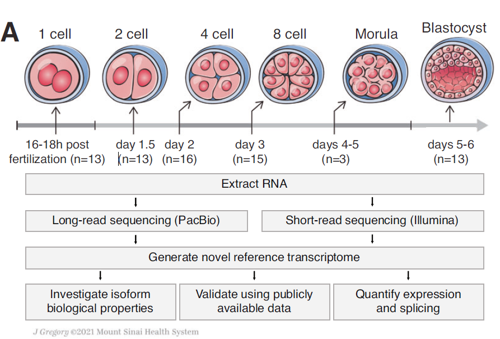
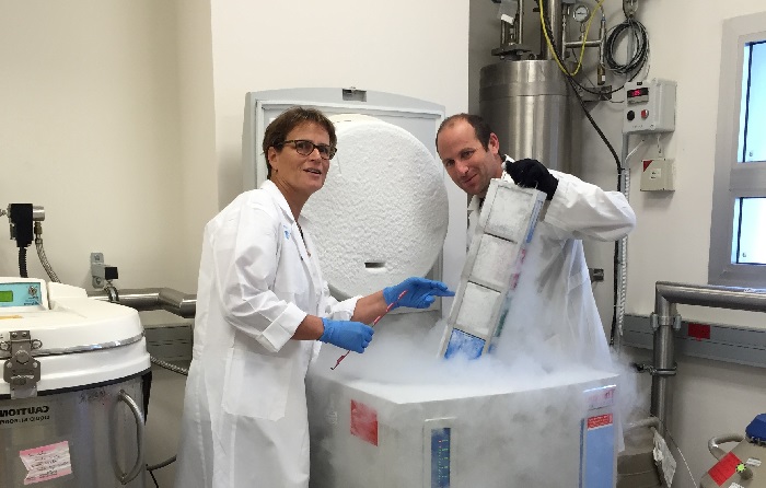
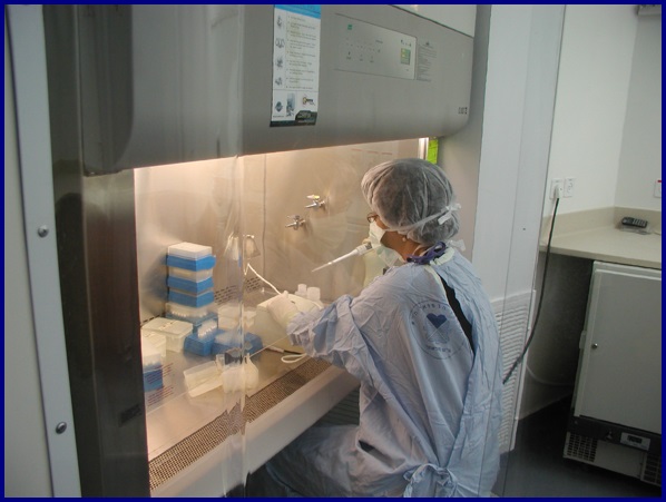
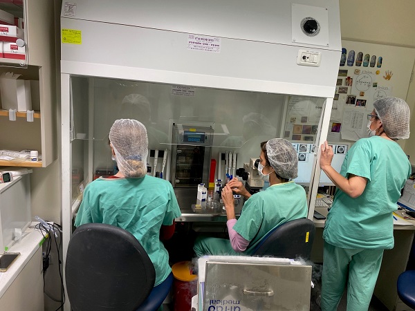
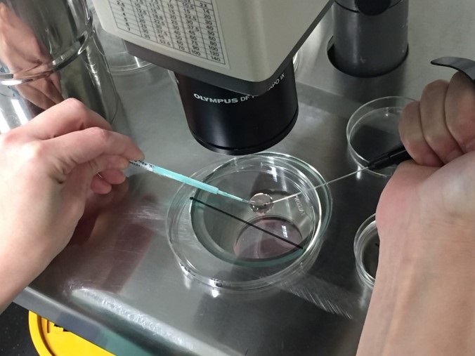
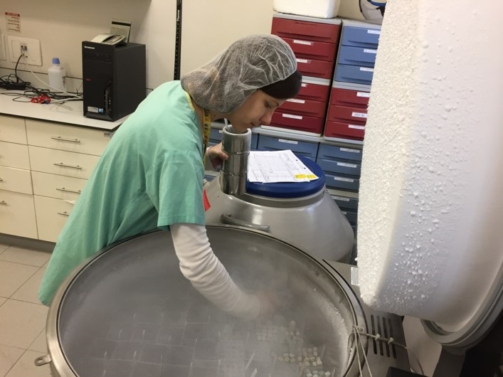
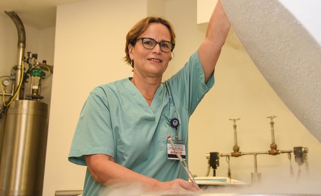
Lab Gallery
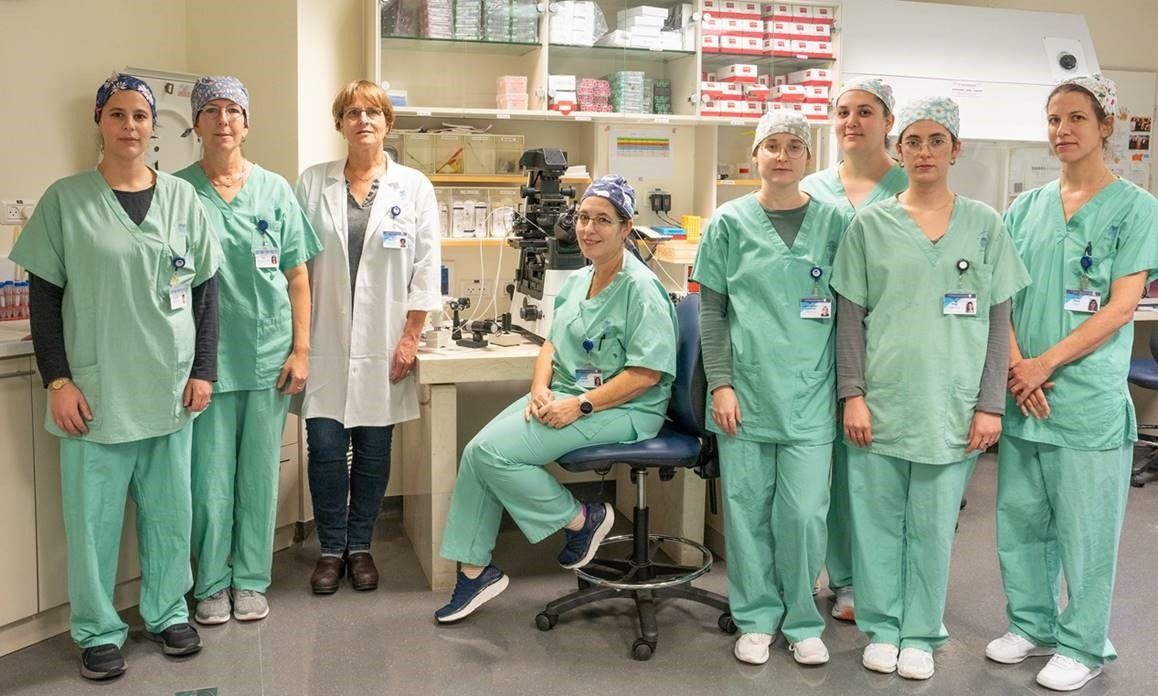
Our Team
Full list of Research Publications
Director; IVF Lab and Wolfe PGD Stem Cell LabRacine IVF Unit; Lis Maternity Hospital
dalitb@tlvmc.gov.il
